Showing posts with label Red Blood Cells. Show all posts

Tuesday, December 22, 2015

The Role of 2,3-Diphophoglycerate


The Role of 2,3-Diphophoglycerate

     The major function of the hemoglobin molecule is the transport of oxygen to the tissues. The oxygen affinity of the hemoglobin molecule is associated with the spatial rearrangement of the molecule and is regulated by the concentration of phosphates, particularly 2,3-DPG in the erythrocyte. The manner in which 2,3-DPG binding to reduced hemoglobin (deoxyhemoglobin) affects oxygen affinity is complex. Basically, 2,3-DPG combines with the beta chains of deoxyhemoglobin and diminishes the molecule’s affinity for oxygen. 


    When the individual heme groups unload oxygen in the tissues, the beta chains are pulled apart. This permits the entrance of 2,3-DPG and the establishment of salt bridges between the individual chains. These activities result in a progressively lower affinity of the molecule for oxygen. With oxygen uptake in the lungs, the salt bonds are sequentially broken; the beta chains are pulled together, expelling 2,3-DPG; and the affinity of the hemoglobin molecule for oxygen progressively increases.
     In cases of tissue hypoxia, oxygen moves from hemoglobin into the tissues, and the amount of deoxyhemoglobin in the erythrocytes increases. This produces the binding of more 2,3-DPG, which further reduces the oxygen affinity of the hemoglobin molecule. If hypoxia persists, depletion of free 2,3-DPG leads to increased production of more 2,3-DPG and a persistently lowered affinity of the hemoglobin molecule for oxygen.


Figure: Hemoglobin molecular changes


Thursday, December 10, 2015

Iron Distribution


Iron-containing compounds in the body are one of two types:
  1. Functional compounds that serve in metabolic (hemoglobin, myoglobin, iron-responsive element-binding protein) or enzymatic (cytochromes, cytochrome oxygenase, catalase, peroxidase) functions
  2. Compounds that serve as transport (transferrin, transferrin receptor) or storage forms (ferritin and hemosiderin) for iron. 
     A poorly understood iron compartment is the intracellular “labile pool.” Iron leaves the plasma and enters the intracellular fluid compartment for a brief time before it is incorporated into cellular components (heme or enzymes) or storage compounds. This labile pool is believed to be the chelatable iron pool. The total iron concentration in the body is 40–50 mg of iron/kg of body weight. Men have higher amounts than women.

    Iron is found primarily in erythrocytes, macrophages, hepatocytes, and enterocytes (absorptive cells at the luminal [apical] surface of the duodenum). Hemoglobin constitutes the major fraction of body iron (functional iron) with a concentration of 1 gm iron/kg of erythrocytes, or about 1 mg iron/mL erythrocytes. Iron in hemoglobin remains in the erythrocyte until the cell is removed from the circulation. Hemoglobin released from the erythrocyte is then degraded in the macrophages of the spleen and liver, releasing iron. Approximately 85% of this iron from degraded hemoglobin is promptly recycled from the macrophage to the plasma where it is bound to the transport protein, transferrin, and delivered to developing normoblasts in the bone marrow for heme synthesis. The macrophages recycle 10 to 20 times more iron than is absorbed in the gut. This iron recycling provides most of the marrow’s daily iron requirement for erythropoiesis.

      Iron in hepatocytes and intestinal enterocytes is stored and utilized as needed to maintain iron homeostasis. The hepatocytes store iron that can be released and utilized when the amount of iron in the plasma is not sufficient to support erythropoiesis. Enterocytes that absorb dietary iron can either export it to the plasma or store it. Iron stored in enterocytes is lost when the cells are sloughed into the intestine. 


Tuesday, December 1, 2015

Glycosylated Hemoglobin


     HbA1C on chromatography is a minor component of normal adult hemoglobin (HbA) that has been modified posttranslationally (HbA3 on starch block electrophoresis). A component usually has been added to the N terminus of the β-chain. The most important subgroup of HbA1 is HbA1C , which has glucose irreversibly attached. This hemoglobin is referred to as glycosylated hemoglobin. HbA1C is produced throughout the erythrocyte’s life, its synthesis dependent on the concentration of blood glucose. Older erythrocytes typically contain more HbA1C than younger erythrocytes having been exposed to plasma glucose for a longer period of time. However, if young cells are exposed to extremely high concentrations of glucose ( >400 mg/dL)  for several hours, the concentration of HbA1C increases with both concentration and time of exposure.
     Measurement of is routinely used as an indicator of control of blood glucose levels in diabetics because it is proportional to the average blood glucose level over the previous two to three months. 
     Average levels of HbA1C are 7.5% in diabetics and 3.5% in normal individuals.


Structure of Hemoglobin


     Hemoglobin is the life-giving substance of every red blood cell, the oxygen-carrying component of the red blood cell. Each red blood cell is nothing more than a fluid-filled sac, with the fluid being hemoglobin. Every organ in the human body depends on oxygenation for growth and function, and this process is ultimately controlled by hemoglobin. In 4 months (120 days), red blood cells with normal hemoglobin content submit to the rigors of circulation. Red blood cells are stretched, twisted, pummeled, and squeezed as they make their way through the circulatory watershed.
The hemoglobin molecule consists of two primary structures:

1- Heme
This structure involves four iron atoms in the ferrous state (Fe2+ ), because iron in the ferric state (Fe3+ ) cannot bind oxygen, surrounded by protoporphyrin IX, or the porphyrin ring, a structure formed in the nucleated red blood cells. Protoporphyrin IX is the final product in the synthesis of the heme molecule. It results from the interaction of succinyl coenzyme A and delta-aminolevulinic acid in the mitochondria of the nucleated red blood cells. Several intermediate by-products are formed, including porphobilinogen, uroporphyrinogen, and coproporphyrin. When iron is incorporated, it combines with protoporphyrin to form the complete heme molecule. Defects in any of the intermediate products can impair hemoglobin function.
2- Globin
This structure consists of amino acids linked together to form a polypeptide chain, a bracelet of amino acids. The most predominant chains for adult hemoglobins are the alpha and beta chains. Alpha chains have 141 amino acids in a unique arrangement, and beta chains have 146 amino acids in a unique arrangement. The heme and globin portions of the hemoglobin molecule are linked together by chemical bonds.
2,3-Diphosphoglycerate (2,3-DPG)
2,3-DPG is a substance produced via the Embden-Meyerhof pathway during anaerobic glycolysis. This structure is intimately related to oxygen affinity of hemoglobin. As 2,3-DPG increased, the affinity of hemoglobin to oxygen is decreased. 

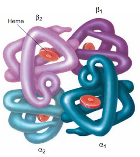
     Each hemoglobin molecule consists of four heme molecules with iron at the center and two pairs of globin chains. The heme structure sits lodged in the pocket of the globin chains. Hemoglobin begins to be synthesized at the polychromatic normoblast stage of red blood cell development. This synthesis is visualized by the change in cytoplasmic color from a deep blue to a lavender-tinged cytoplasmic color. Of hemoglobin, 65% is synthesized before the red blood cell nucleus is extruded, with an additional 35% synthesized by the reticulocyte stage. Normal mature red blood cells have a full complement of hemoglobin, which occupies a little less than one-half of the surface area of the red blood cell.


Figure: Hemoglobin molecule: note four heme molecules tucked inside globin chains.